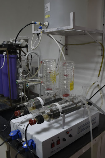
It purifies water by removing more than 99.9% of contaminants, including chemicals, heavy metals, microorganisms, and sediment, via double distillation process. It consists of a boiling chamber, a cooling system, and a separate storage tank. Contact: Prof. Ramesh Sonti.
Category: Biology
.
Biosafety Levels-2 (BSL-2) Facility
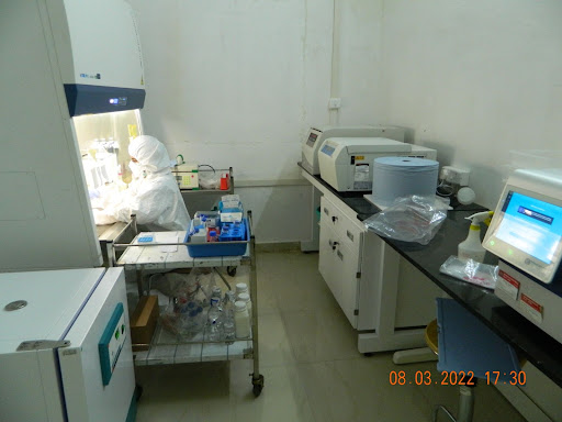
The BSL2 plus facility is used to culture avirulent strains of Mycobacterium tuberculosis and is equipped with a biosafety cabinet, centrifuge, incubator, and plate reader. Contact: Dr. Raju Mukherjee
Plant Tissue Culture and Plant growth room facility
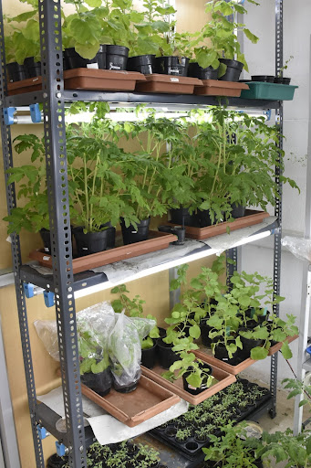
This facility is mainly used for the regeneration of plants using tissue culture based methods. It has a biosafety cabinet, and tissue culture Rack with a digital photoperiod timer. Contact: Dr. Eswarayya Rami Reddy, Dr. Swarup Roy Choudhury, Dr. Annapurna Devi Allu.
Animal Cell Culture Facility
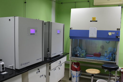
Animal cell culture is used for culturing, and sub-culturing of different types of animal cells. This facility has Bio Safety cabinets, CO2 Incubators, a cell counter Machine, a Centrifugation machine, and a bright field inverted microscope. Contact: Dr. Sajay Kumar, Dr. Vasudharani Devanathan, Dr. Sivakumar Vallabhapurapu
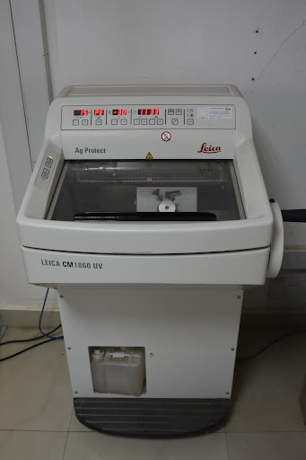
Microtome (cryostat)
For histopathological studies, this device freezes samples to low temperature (upto -35 ℃) to obtain tissue sections ranging from 1 to 100 μm thickness. This device bypass the need of embedding material prior to sectioning of samples. Contact person: Dr. Vasudharani Devanathan
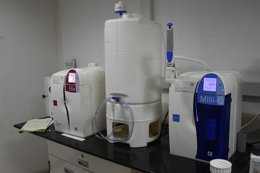
Ultrapure Water Purification Systems
To generate ultra pure water (Type 1 water) of 18.2 MΩ.cm resistance to be used in molecular Biology applications such as PCR, sequencing and for mammalian cell culture. Type 2 water is also generated from the system used in buffer preparation. Contact: Dr. Suchi Goel
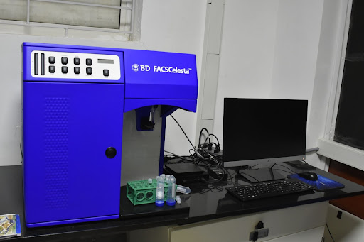
Multicolour Flow Cytometer
The BD FACS Celesta can rapidly and accurately quantify and analyze many kinds of cells, including animal cells, yeast cells, plant cells etc. Contact: Dr. Sivakumar Vallabhapurapu
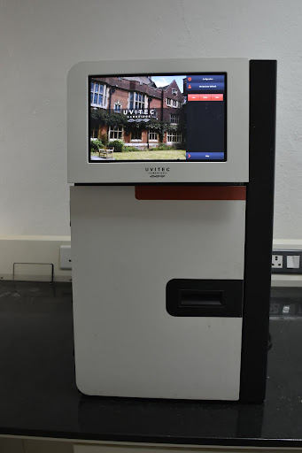
Gel Imaging System Make:UVITEC, Cambridge Model: UVI Doc HD5
Gel Imaging or Gel Documentation system is used to capture digital images of electrophoresis agarose gels in order to obtain a visual record of the experimental results. Contact: Dr. Eswarayya Rami Reddy
Real-Time PCR Systems Model No: CFX Connect Thermal Cycler (96 well and 384 well plates) Make: Bio-Rad Laboratories, USA.
This Polymerase Chain Reaction (PCR) machine is used to monitor the amplification of a targeted DNA molecule during the PCR (in real time), not at its end, as in conventional PCR. Contact: Dr. Sivakumar Vallabhapurapu, Dr. Suchi Goel.
Preprint highlight: DNA damage signaling regulates cohesion stabilization and promotes meiotic chromosome axis morphogenesis
AUTHOR’S – Subramanian, V. V. (2022). Molecular Biology of the Cell, 33(8). DOI: 10.1091/mbc.P22-04-1005


